Image texte

Aim 1. Imaging cell death and inflammation

Marlène Wiart is in charge of the axis.

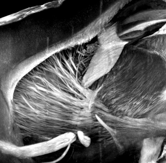
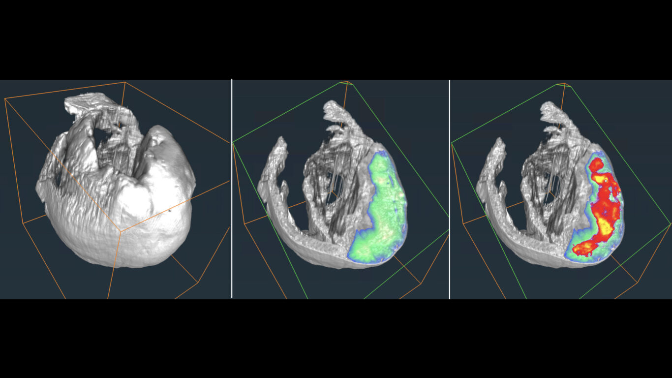
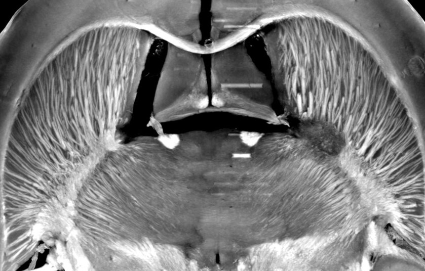
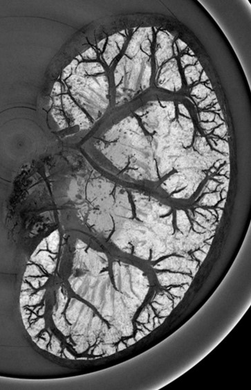
The management of ischemic stroke patients has been revolutionized in the last 5 years with the advent of mechanical thrombectomy, in complement to thrombolysis, to de-obstruct the arteries. This significant therapeutic progress should not hide the fact that ischemic stroke remains a very debilitating pathology: only 45% of thrombectomized patients are independent in their daily life at 3 months. Moreover, 30 to 50% of thrombectomy patients still have poor clinical outcome despite a successful recanalization, the so-called “futile recanalization”. There is therefore an urgent need to propose neuroprotection strategies in order to protect the brain from ischemia and reperfusion damages. To date, none of the therapeutic approaches validated in the pre-clinical arena have been translated into the clinics. One of the reasons for this failure of translational research is a complex physiopathological cascade that is still not completely understood, but also the preclinical methods used are not entirely transposable. We here propose to reveal the determinants of ischemia-reperfusion injuries via molecular imaging tools that characterize and monitor tissue damage in a longitudinal and individual manner. To this aim, we build upon our long-term expertise for multimodal (MRI, PET, CT, US) imaging of edema, perfusion, metabolism and inflammation, in particular macrophage imaging and blood brain barrier dysfunctional. The challenge is to better evaluate and select the treatments to be tested in clinical trials, using a robust and translational methodology. Developing (diagnostic and theranostic) companions imaging tools and their associated imaging biomarkers should favor the clinical transfer of our findings. These imaging tools may further be extended to ischemia/reperfusion syndromes occurring in other organs, such as the heart and the kidney. In conclusion, our ambition is to contribute bridging the gap between pre-clinical and clinical research through in vivo molecular imaging, with the hope, at term, to foster a personalized patient management.


Image texte

One of the reasons for this failure of translational research is a complex physiopathological cascade that is still not completely understood, but also the preclinical methods used are not entirely transposable.
We here propose to reveal the determinants of ischemia-reperfusion injuries via molecular imaging tools that characterize and monitor tissue damage in a longitudinal and individual manner. To this aim, we build upon our long-term expertise for multimodal (MRI, PET, CT, US) imaging of edema, perfusion, metabolism and inflammation, in particular macrophage imaging and blood brain barrier dysfunctional.

The challenge is to better evaluate and select the treatments to be tested in clinical trials, using a robust and translational methodology. Developing (diagnostic and theranostic) companions imaging tools and their associated imaging biomarkers should favor the clinical transfer of our findings.
These imaging tools may further be extended to ischemia/reperfusion syndromes occurring in other organs, such as the heart and the kidney. In conclusion, our ambition is to contribute bridging the gap between pre-clinical and clinical research through in vivo molecular imaging, with the hope, at term, to foster a personalized patient management.

Image texte

Aim 2. Molecular mechanisms of ischemia-reperfusion (IR): Ca2+, mitochondria, metabolism and inflammation

Mélanie Paillard is in charge of the axis.

The molecular mechanisms of ischemia-reperfusion injury start to be better elucidated, notably in the heart with a key role of mitochondria in cell death. Our previous works have shown that the mitochondrial permeability transition pore (PTP) opening plays a crucial role in cell death regulation. However, its inhibition in clinics did not afford a beneficial prognostic to patients undergoing myocardial infarct or cardiac arrest. We have thus reconsidered our research on cardioprotection by identifying new molecular targets upstream the PTP, involved notably in Ca2+ exchange between reticulum and mitochondria. Our work is focusing: 1) on the reticular side at Ca2+ channels, more precisely the IP3 receptor, the heat receptor TRPV1, the passive translocon leak channel and the cold receptor TRPM8; and 2) on the mitochondrial side, at the entry of Ca2+ via the calcium uniporter MCU/MICU1 and the PTP regulation. We aim at unraveling the molecular and signaling mechanisms, notably by the GSK3β kinase and the transcription factor STAT3, together with determining the metabolic and inflammatory impact of their alteration or modulation. From molecular analysis to pre-clinical study, our research seeks to propose new therapeutic targets to improve the long-term outcome of patients undergoing an ischemia-reperfusion event either in the heart, brain or kidney.

Image texte

Aim 3. Mechanisms of organ conditioning, preservation & engineering

Gabriel Bidaux is in charge of the axis.

Among the protection strategies against ischemia-reperfusion lesions in pre-clinical or clinical settings, therapeutic hypothermia is applied in post-cardiac arrest resuscitation, neonatal pediatrics, cardiac surgery and renal, and organ preservation. The ischemic conditioning methods: pre- and post-conditioning type, local or remote, have demonstrated protective effects in preclinical models and few promising results in the clinic. However, these methods have mainly been studied empirically and they suffer from the lack of knowledge on the molecular mechanisms involved. This research axis is based on a top-down strategy as well as the use of unsupervised high-throughput approaches but also of innovative targeted approaches (cold receptors) to better describe the mechanisms at work. Our final objective is to be able to optimize clinical protocols using cold as a cytoprotective and therapeutic medium. On the other hand, we are developing innovative solutions to optimize the preservation of grafts in deep hypothermia (Inovagraft, INSERM 2015 innovation prize), as well as the follow-up of patients in post-cardiac arrest resuscitation in therapeutic hypothermia.

Within this multidisciplinary and technology-oriented research axis, we also collaborate on the development of new systems and technologies to improve early diagnosis, post-reperfusion follow-up of patients at hospital and of innovative systems to better study our animal models.

Image texte
Cardiology

Cardiology

Cardiovascular diseases are the main cause of death in France among women, and the second cause for men, representing more than 120,000 patients per year (Santé Publique France). Myocardial infarction is caused by ischemia of the heart muscle. If the recent improvement in the management of myocardial infarction, and in particular systematic reperfusion (coronary angiography and angioplasty) has made it possible to reduce its mortality (15,000 deaths per year in France), secondary events such as insufficiency chronic heart disease (20 to 25% of patients after a heart attack) or ischemic recurrence remain high and are the estimated cause of 70,000 annual deaths. The larger the size of the infarction, the more numerous and severe the complications will be. Our clinical research themes focus on: the search for early post-infarction prognostic biomarkers (in order to target the population at risk of presenting complications in the context of the development of personalized medicine) and the search for treatments to reduce the size infarction, to improve left ventricular remodeling and, ultimately, to reduce post-infarction morbidity and mortality (cardioprotective treatments).


Cardiac arrest kills around 40,000 people a year in France. Its mortality is very high (92%) and few therapeutic advances have been made in recent years. Among post-cardiac arrest treatments, therapeutic hypothermia occupies an important place thanks to supposed neuroprotective effects. With this in mind, we are developing clinical trials to assess the effects of therapeutic hypothermia in patients who have presented with cardiac arrest, but also fundamental work to better understand the molecular mechanisms at work. We are also working to find prognostic markers that would allow us to adjust the treatment to each patient.


Cardiac surgery (aorto-coronary bypasses, replacement of valves, replacement of the ascending aorta, etc.), when performed with the heart stopped (under extracorporeal circulation = CEC) is subject to the deleterious effects of ischemia-reperfusion. Appropriate cardioplegia can partially prevent these deleterious effects. We are working to develop new cardioprotection strategies during cardiac surgery under CEC as well as to precisely describe the intracellular mechanisms responsible for myocardial damage during CEC.

Image texte
Neurology

Neurology

Cerebrovascular accidents (CVA) are a major source of disability and are in 80% of cases of ischemic nature, linked to an arterial obstruction. Intravenous thrombolysis and more recently thrombectomy, which consists of mechanically unclogging the artery, make it possible to reduce the neurological handicap. However, even with arterial opening, brain damage may worsen due to secondary damage, related in part to the intensity of the post-ischemic inflammatory reaction. To date, the understanding of these mechanisms remains partial, and may limit the efficacy of neuroprotective strategies.

In order to better understand these phenomena, we set up the HIBISCUS-STROKE cohort (CoHort of Patients to Identify Biological and Imaging markerS of CardiovascUlar Outcomes in Stroke) including the patients treated by thrombectomy for an ischemic stroke in the Neurovascular Unit of Pierre Wertheimer Neurological Hospital (Lyon, France) , in the context of RHU MARVELOUS (ANR-16-RHUS-0009). These patients are being longitudinally assessed for blood markers of inflammatory response and ischemic injury using magnetic resonance imaging.

The objective of our work is to better characterize the mechanisms of the post-ischemic inflammatory response and to specify the neuroprotection strategies. This work has already been the subject of several publications in international peer-reviewed journals.

Image texte
Nephrology

Nephrology

Kidney disease now affects 5 to 10% of French people and is associated with significant cardiovascular morbidity and mortality. Acute renal failure results from transient and most often reversible kidney dysfunction caused by an ischemic or hemorrhagic event, sepsis, or taking nephrotoxic drugs. Conversely, chronic renal failure results from progressive but irreversible destruction of the kidneys. In about 50% of cases, chronic kidney disease is the result of diabetes or high blood pressure. In France, end-stage renal disease affects more than 89,692 people (REIN Network 2018) and requires the use of dialysis or kidney transplantation to ensure patient survival.


The objectives of our project are therefore: (1) to better understand the pathophysiology of ischemia / renal reperfusion damage; (2) to get a better comprehension of the mechanisms involved in the occurrence of uremic heart disease and the increased cardiovascular risk in a situation of renal failure; (3) to evaluate new strategies for organ protection and / or conservation such as therapeutic hypothermia and (4) to assess renal performance using functional imaging.


Donate to research

We are developing preventive strategies and new treatments for better patient care. Researchers are counting on you!

Support research